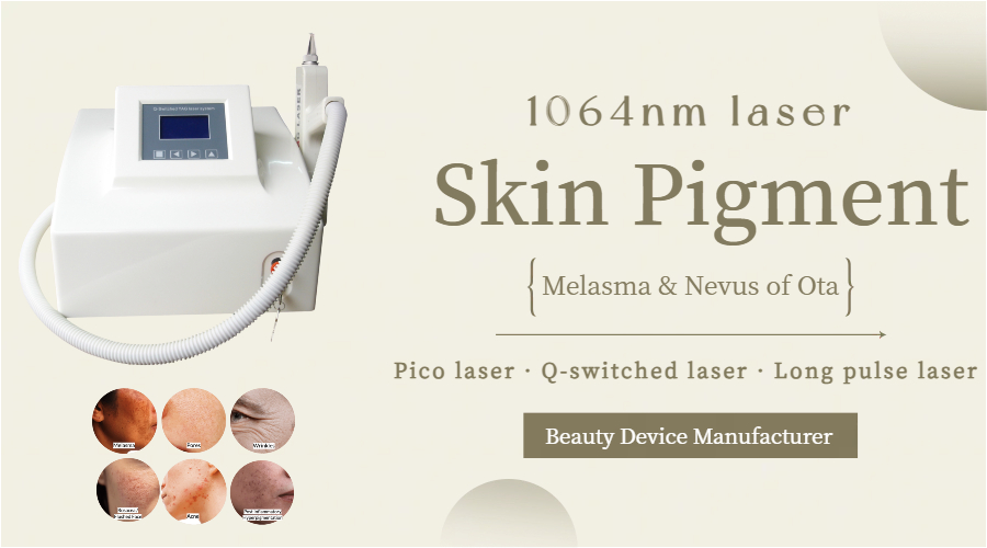
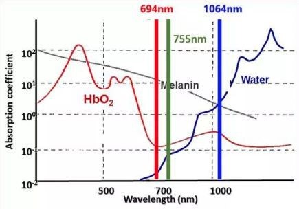
1064 nm wavelength laser belongs to a special band of infrared light. According to the selective photothermal characteristics of skin color base, 1064 nm wavelength laser can be absorbed by water, hemoglobin and melanin in the skin at the same time.
However, the absorption of 1064 nm by the three target color bases is relatively weak. The collagen fibers and adipose tissue in the dermis can also absorb this wavelength of light. Studies have found that the penetration depth of 1064 nm laser in skin tissue can reach 5~10mm. The same wavelength of 1064 nm laser can adjust the energy and pulse width to make the depth of action from epidermis to dermis, then to shallow fat and deep blood vessels, and solve skin pigmentation diseases, vascular diseases and various skin problems in a targeted manner.
This article focuses on the clinical application of 1064 nm laser in skin pigmentation diseases.
1064nm can be distinguished based on different pulse widths.
1064 nm picosecond laser.
Picosecond pulse width. Through photothermal and photomechanical effects, it can effectively act on pigment particles. Picosecond laser can be used for tattoos, pigment diseases and facial rejuvenation, and has certain effects. Picosecond laser, which has advantages in pulse width, has gradually replaced nanosecond laser in the treatment of pigmentary diseases. It can not only improve the treatment effect of pigmentary diseases, but also has advantages such as short treatment cycle and mild adverse reactions for those who are ineffective with nanosecond laser treatment. And complications such as pain, edema, transient erythema, and scab are milder than nanosecond laser.
1064 nm Q-switched laser.
Nanosecond pulse width is the preferred choice for the treatment of chloasma, nevus of Ota, brown-blue nevus and other pigmentary abnormal skin diseases in deeper parts. Retrospective studies have shown that there is no significant difference in the effectiveness of picosecond laser and Q-switched laser in the treatment of patients with dark skin and pigmentary diseases such as Baker nevus, nevus of Ito, and melanosis.
1064 nm long pulse width laser.
The microsecond pulse width can cause thermal damage to target cells, and the pulse width parameters are adjustable. The product is applicable to a wide clinical range and has high treatment safety and comfort.
Clinical application of 1064nm wavelength laser in skin pigmentation diseases.
First, we need to understand the melanin distribution depth of common pigmentary diseases:
| Melanin distribution depth |
Common pigmentary diseases |
| Epidermal type | Freckles; freckles; coffee-coffee spots; solar lentigo; seborrheic keratosis |
| Epidermal-dermal type | Melasma; Baker’s nevus; nevus maculatus; post-inflammatory pigmentation, etc. |
| Leather type | Nevus of Ota; Nevus of Ito; Tattoo; Brown-blue nevus on the cheekbone, etc. |
Some epidermal spots are more suitable for treatment with 1064nm laser than 532nm.
When laser is used clinically to treat pigmentary diseases, the laser wavelength can usually be selected according to the different depths and colors of the skin lesions. At present, Q-switched laser is mostly used to treat epidermal pigmented lesions. It is highly selective for the treatment of freckles, freckle-like nevi, and coffee spots, and is currently the best treatment method.
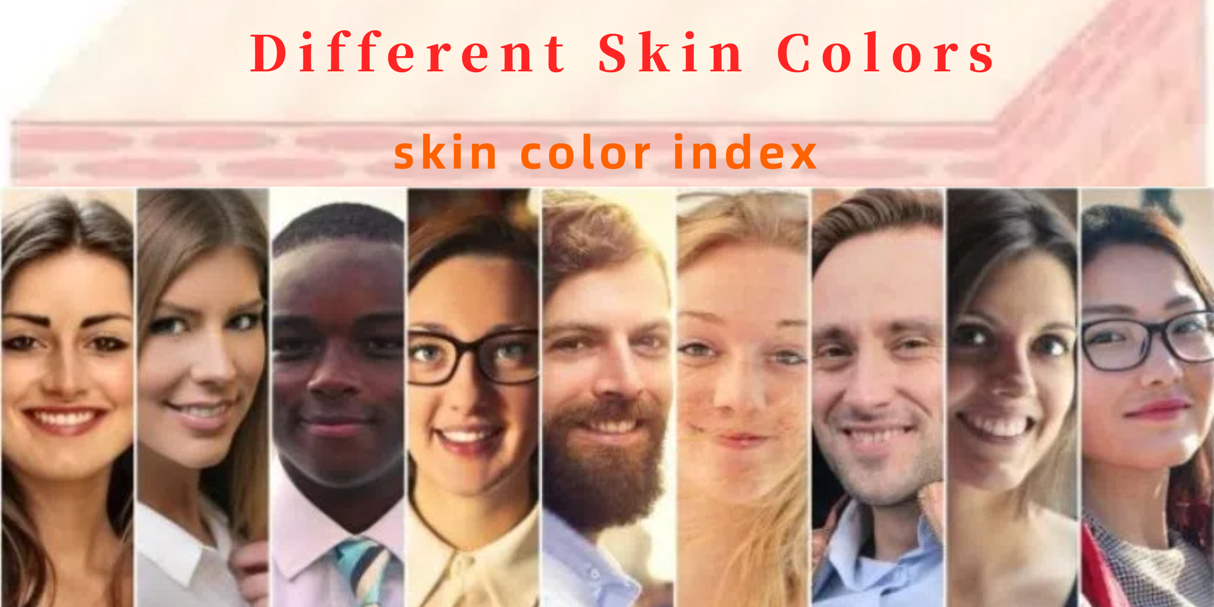
However, the choice of wavelength is relative rather than absolute. In Asian populations, the shorter wavelength 532/694/755nm laser treatment of pigmentary diseases takes the appearance of ice white phenomenon (IWP) as the treatment endpoint. After irradiation, the lesion site turns white instantly. After surgery, redness, swelling, pain, scabs, bleeding, small blisters, blood blisters and other adverse reactions may occur. After the scab falls off, post-inflammatory hyperpigmentation (PIH) is prone to occur. PIH is more likely to occur in individuals with Fitzpatrick skin type III to VI, usually with 3 to 12 months or longer. When PIH occurs, it is not recommended to immediately perform the next freckle removal laser treatment until PIH subsides, which will also prolong the treatment course of epidermal spots.
The end point of 1064nm laser is the slight erythema of the skin, and the treatment purpose can be achieved without frost whitening. Although it requires more treatments than short-wavelength laser, the final result shortens the adverse reaction period and reduces pigmentation and pigmentation. Probability of depigmentation. Therefore, 1064nm laser is a good choice for dark-skinned people and patients with epidermal plaques who have special requirements for side effects and downtime after laser surgery and who are intolerant to pain. Studies have proven that Q-switched 1064nm Nd:YAG laser is effective in treating café-au-lait spots and sunspots.
During treatment, the treatment parameters can be initially selected according to the patient’s age, skin color, texture, and sensitivity. For the first treatment, a low energy density suitable for the patient is adjusted in the hidden skin lesion area. The laser handle is vertically aligned with the lesion, and the light spot is irradiated onto the skin surface in front of the ear. A quick scan is performed to observe the local reaction to determine the energy density, and then the lesion is scanned multiple times to determine the degree of skin flushing. Cold compress is applied immediately after the treatment. The treatment frequency is once every 2 to 3 weeks, 5 to 8 times, and follow-up is 6 months after the last treatment.
For “difficult” mixed pigmentary diseases, 1064nm has unique advantages.
For pigmentary diseases with lesions at the junction of the true epidermis, such as melasma and post-inflammatory pigmentation, the pathological manifestations are an increase in melanophages, accompanied by inflammation and systemic or local pigment metabolism abnormalities. Such pigmented lesions require extra caution when using laser treatment. Currently, the recommended laser for the treatment of melasma is a large-spot, low-energy Q-switched 1064 nm Nd:YAG laser (the recommended parameters are a spot diameter of ≥6 mm and an energy of ≤3 J/cm2). The large-spot, low-energy Q-switched laser can selectively destroy melanosomes in melanocytes, minimize damage to surrounding skin tissue and basement membranes, and avoid the occurrence of postoperative inflammatory pigmentation. The Q-switched 1064 nm Nd:YAG laser has also been shown to have good efficacy in some PIH cases.
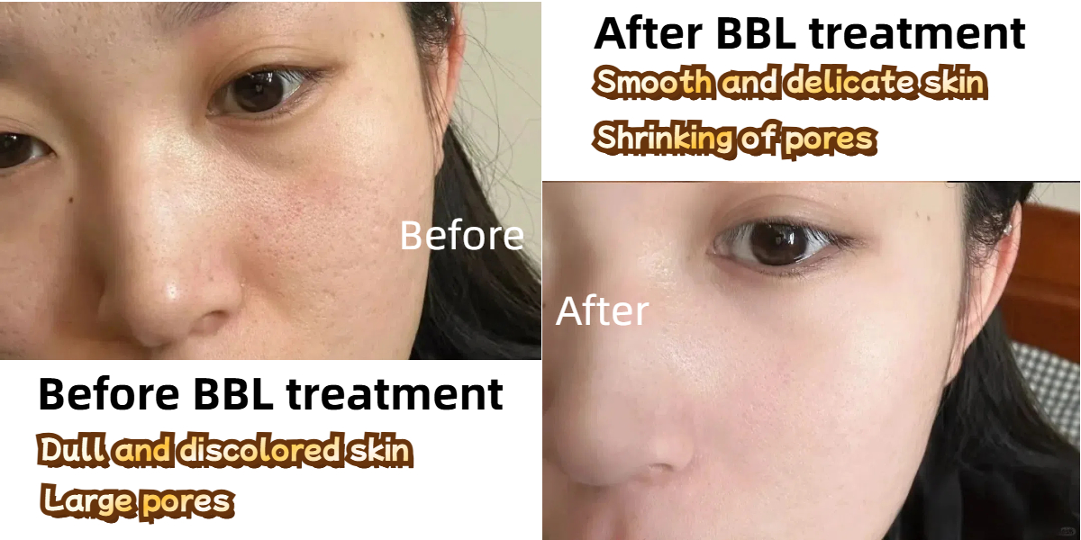
In a clinical study involving 78 patients with post-acne pigmentation, the patients received 6 treatments of 1064nm laser (2 weeks/interval), and clinical photos showed that 70% of the patients had significant improvement in pigmentation. Compared with 694nm laser, low-energy 1064nm laser penetrates deeper and can target low energy to melanin particles in keratinocytes, melanocytes, and skin melanin phagosomes. Since melanin absorbs less energy, it is relatively safe in the treatment of PIH in patients with dark skin.
For pigmented diseases that penetrate deep into the dermis, 1064nm has an absolute advantage.
The pigment deposition site in dermal hyperpigmented lesions is deeper. The abnormal melanocytes of Ota nevus are distributed in the papillary layer and the shallow reticular layer of the dermis. The pathology of the zygomatic brown-blue nevus is similar to that of Ota nevus, but the location of abnormal pigment cells is usually more superficial than that of Ota nevus. Because the pigment of this type of disease is deep, and the number and size of mature melanosomes in the dermis are larger than those in the epidermis, a Q-switched laser with a wavelength of 1064 nm should be selected for treatment, and the energy density should be appropriately increased. Although a small amount of energy will be absorbed by the epidermal melanin shunt, there is still enough energy to enter the dermis, causing the disintegration and fragmentation of the pigment bodies in the dermis. The pigment particle debris is phagocytosed by macrophages and then excreted from the body through the blood and lymphatic circulation, which selectively removes the pigment particles or pigment cells in the dermis to the greatest extent and reduces the damage to adjacent normal cells. Similarly, when treating patients with dark skin, attention should be paid to appropriately reduce the energy density to avoid PIH.
The endpoint of treatment of Ota nevus using 1064 nm Nd:YAG laser is when the skin in the treatment area turns white and a small amount of bleeding appears after a few minutes.
The higher the energy density, the better the corresponding treatment effect. However, too high energy density may damage the surrounding normal tissues, thus causing scars. If blisters or intensive bleeding form at the treatment site, the energy density should be reduced. For skin lesions with shallow lesions, large light spots and low energy density can be used for treatment; for skin lesions with varying depths or deep into the subcutaneous tissue, small light spots and high energy density should be used for treatment.
The zygomatic brown-blue nevus is a common pigmented skin disease in young and middle-aged women. During treatment, the appropriate light spot and energy density should be selected according to the patient’s age, skin color type, lesion site and characteristics. If the treatment energy is too large, pigmentation is prone to occur. If the energy is too small, the pigmentation will not disappear significantly, and the number of treatments should be increased. It is recommended that a light spot test must be performed before treatment. Some patients’ skin lesions do not have bruises during treatment, but punctate bleeding can be seen after 3 to 5 minutes of observation. The treatment end point is mild bruises. If pigmentation occurs during treatment, it is recommended to continue laser treatment after the pigmentation disappears. In areas where the subcutaneous tissue of the temporal region is thinner, the energy is reduced accordingly to reduce the occurrence of complications.
Picosecond 1064nm laser is more effective in removing black and blue tattoos.
Tattoos, as a personalized body art phenomenon, are quite popular among young people, but there is also an increasing demand for tattoo removal. Since tattoos have many colors and the pigment particles are located deep, some even exceed the dermis, it is difficult to treat.
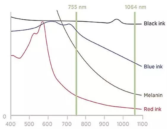
Different pigments have different light wave absorption peaks, so when treating tattoos, it is necessary to choose the appropriate wavelength according to the color of the tattoo dye. 1064nm Nd:YAG laser is mainly used to remove black and blue tattoos.
The short pulse width and high energy of picosecond lasers make it show unique advantages in the application of tattoos. Compared with traditional Q-switched lasers, picosecond lasers have shorter pulse widths and higher energy density. The photomechanical force generated by picosecond lasers breaks the pigment target base into smaller pigment particles, which are easier to be engulfed by phagocytes and excreted from the body through the lymphatic system, effectively improving the clearance rate of pigments and reducing the transfer of heat to surrounding tissues, thereby reducing damage to surrounding tissues and the risk of scar formation and pigmentation.
Summary of 1064 nm laser treatment.
As a unique band of near-infrared light, 1064nm laser can treat epidermal spots in people with dark skin, chloasma and post-inflammatory pigmentation involving the epidermis and dermis, as well as pigmentary diseases with deeper lesions and higher pigment density. It has remarkable effects on various blue and black tattoo treatments.
When applying 1064nm laser for clinical treatment, laser instruments with different pulse widths can be selected according to the type of disease, the location of the disease, and the patient’s personal situation (skin color, age, skin type, etc.), and can be targeted through parameter adjustment of energy and spot size. Effectively solve skin pigment diseases and give full play to the unique advantages of 1064nm laser.
FAQs about 1064 nm laser.
Q: How does the 1064nm laser work?
A: ND YAG laser emits light with a wavelength of 064nm. Since light is penetrating, it can act on different skin layers according to different wavelengths. 1064 has a higher absorption coefficient for pigments and can preferentially absorb pigment cells, thereby treating pigmented skin problems.
Q: What is 1064 nm laser?
A: 1064 nm laser refers to light with a single wavelength of 1064 nm. Laser refers to light of a specific wavelength. By removing other wavelengths and retaining only the 1064 nm wavelength, it becomes a 1064 nm laser.
Q: is 1064 nm laser visible?
A: 1064 nm laser is invisible light. The wavelength range of visible light is between 390 nm and 780 nm, and 1064 nm wavelength is obviously not within the range.
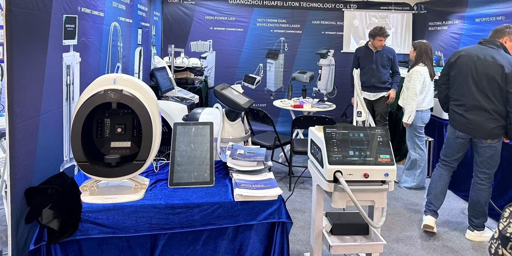
How to buy a 1064 nm laser machine?
LitonLaser is a professional medical equipment manufacturer. We have many laser machines with 1064 nm wavelength, such as Q-switched laser & picosecond laser & 1064 long pulse laser hair removal & diode laser. If you need any 1064nm wavelength laser, please feel free to contact us.